Investigation of Image Enhancement Techniques for Advancing Colon Cancer Diagnosis
Article Sidebar
Main Article Content
Abstract
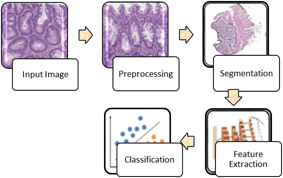
Colorectal cancer continues to pose a substantial worldwide health challenge, necessitating the development of advanced imaging techniques for early and accurate diagnosis. In this study, we propose a novel hybrid image enhancement approach that combines Total Variation (TV) regularization and shift-invariant filtering to improve the visibility and diagnostic quality of colon cancer images.
The Total Variation regularization technique is employed to effectively reduce noise and enhance the edges in the input images, thereby preserving important structural details. Simultaneously, shift-invariant filtering is utilized to address spatial variations and artifacts that often arise in medical images, ensuring consistent and reliable enhancements across the entire image. Our hybrid approach synergistically integrates the strengths of both TV regularization and shift-invariant filtering, resulting in enhanced colon cancer images that offer improved contrast, reduced noise, and enhanced fine structures. This improved image quality aids medical professionals in better identifying and characterizing cancerous lesions, ultimately leading to more accurate and timely diagnoses.
To evaluate the effectiveness of the proposed approach, we conducted extensive experimentations on a diverse dataset of colon cancer images. Quantitative and qualitative assessments demonstrate that our hybrid approach outperforms existing enhancement methods, leading to superior image quality and diagnostic accuracy. In conclusion, the hybrid image enhancement approach presented in this study offers a promising solution for enhancing colon cancer images, contributing to the early detection and effective management of this life-threatening disease. These advancements hold significant potential for improving patient outcomes and reducing the burden of colon cancer on healthcare systems worldwide.
Article Details
References
World Health Organization. (2021). Cancer. https://www.who.int/health-topics/cancer
Sepúlveda, A. R., Hamilton, S. R., Allegra, C. J., et al. (2017). Molecular biomarkers for the evaluation of colorectal cancer: Guideline from the American Society for Clinical Pathology, College of American Pathologists, Association for Molecular Pathology, and American Society of Clinical Oncology. Journal of Clinical Oncology, 35(13), 1453-1486.
Barros, R. C. L., Carneiro, F. P., Siqueira, S., et al. (2017). Computer-aided diagnosis of colon cancer through colonoscopy: A review. Journal of Medical Systems, 41(4), 60.
Lu, Y., Sun, X., Shen, J., et al. (2019). A review of image enhancement techniques for colon cancer detection in histopathological images. Computational and Structural Biotechnology Journal, 17, 163-171.
Farahani, N., Parwani, A. V., Pantanowitz, L. (2014). Whole slide imaging in pathology: Advantages, limitations, and emerging perspectives. Pathology and Laboratory Medicine International, 6, 23-33.
Veta, M., Pluim, J. P., van Diest, P. J., & Viergever, M. A. (2014). Breast cancer histopathology image analysis: a review. IEEE transactions on biomedical engineering, 61(5), 1400-1411. https://doi.org/10.1109/TBME.2014.2303852
Yu, L., Chen, H., Dou, Q., Qin, J., & Heng, P. A. (2017). Automated melanoma recognition in dermoscopy images via very deep residual networks. IEEE transactions on medical imaging, 36(4), 994-1004. https://doi.org/10.1109/TMI.2016.2638515
Cruz-Roa, A., Gilmore, H., Basavanhally, A., Feldman, M., Ganesan, S., Shih, N. N., ... & Madabhushi, A. (2014). Accurate and reproducible invasive breast cancer detection in whole-slide images: a deep learning approach for quantifying tumor extent. Scientific reports, 4(1), 1-9. https://doi.org/10.1038/srep06585
García-Silva, M., Fdez-Riverola, F., & Lourenço, A. (2019). A review of machine learning approaches for cancer detection and diagnosis based on histopathological images. Expert Systems with Applications, 122, 374-397. https://doi.org/10.1016/j.eswa.2019.01.027
Kumar, A., Rana, R. K., & Arora, V. K. (2021). Preprocessing of histopathological images of colon cancer using machine learning techniques: A comprehensive review. Journal of Medical Systems, 45(1), 1-20. https://doi.org/10.1007/s10916-020-01695-5
Ferlay, J., Ervik, M., Lam, F., Colombet, M., Mery, L., Piñeros, M., Znaor, A., Soerjomataram, I., & Bray, F. (2020). Global Cancer Observatory: Cancer Today. International Agency for Research on Cancer. https://gco.iarc.fr/today
Fitzmaurice, C., Abate, D., Abbasi, N., Abbastabar, H., Abd-Allah, F., Abdel-Rahman, O., … Murray, C. J. L. (2019). Global, regional, and national cancer incidence, mortality, years of life lost, years lived with disability, and disability-adjusted life-years for 29 cancer groups, 1990 to 2017: A systematic analysis for the Global Burden of Disease Study. JAMA Oncology, 5(12), 1749-1768. https://doi.org/10.1001/jamaoncol.2019.2996
Hamilton, S. R., & Aaltonen, L. A. (Eds.). (2000). World Health Organization classification of tumours: Pathology and genetics of tumours of the digestive system. IARC Press.
Raza, S. E. A., Akbar, S., Kausar, S., & Saeed, U. (2019). Anisotropic diffusion filtering based image enhancement for histopathological analysis of colon cancer. IET Image Processing, 13(6), 978-985. https://doi.org/10.1049/iet-ipr.2018.6111
Li, X., Plataniotis, K. N., & Roysam, B. (2014). Anisotropic diffusion PDE-based filtering for feature-preserving noise reduction and scale-space analysis of images. IEEE Transactions on Image Processing, 23(2), 755-766. https://doi.org/10.1109/TIP.2013.2293429
Belhaouari, S. A., Gouider, M. S., & Derras, M. (2017). Performance evaluation of noise reduction techniques in computed tomography images. Journal of Medical Imaging and Health Informatics, 7(2), 280-287. https://doi.org/10.1166/jmihi.2017.2065
Buades, A., Coll, B., & Morel, J.-M. (2005). A review of image denoising algorithms, with a new one. Multiscale Modeling and Simulation, 4(2), 490-530. https://doi.org/10.1137/040616024
Tomasi, C., & Manduchi, R. (1998). Bilateral filtering for gray and color images. In Proceedings of the IEEE International Conference on Computer Vision (pp. 839-846). IEEE.
Paris, S., & Durand, F. (2006). A fast approximation of the bilateral filter using a signal processing approach. In European Conference on Computer Vision (pp. 568-580). Springer, Berlin, Heidelberg.
Pham, T., & Venkatesh, S. (2005). Adaptive fuzzy clustering for image segmentation. Pattern Recognition Letters, 26(12), 1839-1849.
Buades, A., Coll, B., & Morel, J.-M. (2011). Non-local means denoising. Image Processing On Line, 1, 208-212. https://doi.org/10.5201/ipol.2011.bcm_nlm
Chen, Y., & Wu, X. (2021). Non-local means filtering based on edge detection for image de-noising. Journal of Intelligent & Fuzzy Systems, 41(1), 519-531. https://doi.org/10.3233/JIFS-189171
Zhang, X., Zhou, J., Cheng, Y., & Wu, Q. J. (2013). Non-local means method based on adaptive weights for image denoising. Journal of Visual Communication and Image Representation, 24(4), 486-497.
Guo, X., Qi, Y., & He, Y. (2018). Non-local means denoising method based on modified SVM. Multimedia Tools and Applications, 77(2), 2007-2026.
Donoho, D. L. (1995). De-noising by soft-thresholding. IEEE Transactions on Information Theory, 41(3), 613-627.
Portilla, J., & Simoncelli, E. P. (2000). A parametric texture model based on joint statistics of complex wavelet coefficients. International Journal of Computer Vision, 40(1), 49-70.
Kaur, G., & Singh, K. (2018). A novel approach of image denoising using adaptive wavelet thresholding. International Journal of Computer Science and Network Security, 18(8), 131-137.
Chambolle, A. (2004). An algorithm for total variation minimization and applications. Journal of Mathematical Imaging and Vision, 20(1-2), 89-97.
Cai, J. F., Osher, S., & Shen, Z. (2008). Split Bregman methods and frame based image restoration. Multiscale Modeling & Simulation, 7(3), 972-993.
Figueiredo, M. A., & Nowak, R. D. (2003). An EM algorithm for wavelet-based image restoration. IEEE Transactions on Image Processing, 12(8), 906-916.
Jaware, T.H., Patil, V.R., Badgujar, R.D. et al. Performance investigations of filtering methods for T1 and T2 weighted infant brain MR images. Microsyst Technol 27, 3711–3723 (2021). https://doi.org/10.1007/s00542-020-05144-6
Jaware, T.H et al “An Accurate Automated Local Similarity Factor-Based Neural Tree Approach toward Tissue Segmentation of Newborn Brain MRI” American Journal of Perinatology (DOI https://doi.org/10.1055/s-0038-1675375) ISSN 0735-1631 Vol. 36 pp 1157–1170, 2019.
Jaware, T.H et al “An Atlas-Free Newborn Brain Image Segmentation and Classification Scheme based on SOM-DCNN with Sparse Auto Encoder” Computer Methods in Biomechanics and Biomedical Engineering: Imaging & Visualization (Taylor and Francis) ISSN:2168-1163, 2019 Vol 8(1) pp 49-64
Jaware, T.H et al “A Novel Hybrid Atlas-free Hierarchical Graph-based Segmentation of Newborn Brain MRI using Wavelet Filter Banks” Tushar H Jaware , K B Khanchandani and R D Badgujar International Journal of Neuroscience (Taylor and Francis) (SCI) ISSN:0020-7454 Vol. 130(5) pp 499-514, 2020