Detection of Pulmonary Embolism: Workflow Architecture and Comparative Analysis of the CNN Models
Article Sidebar
Main Article Content
Abstract
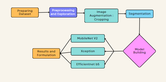
Machine learning has proven to be a practical medical image processing technique for pattern discovery in low-quality labelled and unlabeled datasets. Deep vein thrombosis and pulmonary embolism are both examples of venous thromboembolism, which is a key factor in patient mortality and necessitates prompt diagnosis by experts. An immediate diagnosis and course of treatment are necessary for the life-threatening cardiovascular condition known as pulmonary embolism (PE). In the study of medical imaging, especially the identification of PE, machine learning (ML) algorithms have produced encouraging results. This study's objective is to assess how well machine learning (ML) algorithms perform in identifying PE in computed tomography (CT) scans. A range of ML approaches were used to the dataset, including deep learning algorithms such as convolutional neural networks. The effectiveness of PE detection systems can be greatly enhanced by the use of cutting-edge methodologies like deep learning, which lowers the possibility of incorrect diagnoses and enables the quick administration of therapy to individuals who require it. This work contributes to the growing body of evidence that supports the use of ML in medical imaging and diagnosis. Future research should examine how these algorithms might be included into clinical workflows, resolving any potential implementation challenges, and making sure their adoption is done so in a secure and efficient way. In this study, we provide a thorough evaluation of three different models: the streamlined architecture MobileNetV2 with an accuracy of 96%, compared to other models like the Xception model with an accuracy of 91%, and the Efficientnet B5 model with an accuracy of 97%, after observation and process following.
Article Details
References
A. K. Tarbox and M. Swaroop, “Pulmonary embolism,” Int J Crit Illn Inj Sci, vol. 3, no. 1, p. 69, 2013, doi: 10.4103/2229-5151.109427.
X. Yang et al., “SPECIAL SECTION ON DATA-ENABLED INTELLIGENCE FOR DIGITAL HEALTH A Two-Stage Convolutional Neural Network for Pulmonary Embolism Detection From CTPA Images”, doi: 10.1109/ACCESS.2019.2925210.
M. L. Giger, “Machine Learning in Medical Imaging,” Journal of the American College of Radiology, vol. 15, no. 3, pp. 512–520, Mar. 2018, doi: 10.1016/J.JACR.2017.12.028.
D. Shen, G. Wu, and H.-I. Suk, “Deep Learning in Medical Image Analysis,” 2017, doi: 10.1146/annurev-bioeng-071516.
H. Khachnaoui, M. Agrebi, S. Halouani, and N. Khlifa, “Deep Learning for Automatic Pulmonary Embolism Identification Using CTA Images,” in 2022 6th International Conference on Advanced Technologies for Signal and Image Processing (ATSIP), May 2022, pp. 1–6. doi: 10.1109/ATSIP55956.2022.9805929.
R. Pillai, P. Oza, and P. Sharma, “Review of Machine Learning Techniques in Health Care,” 2020, pp. 103–111. doi: 10.1007/978-3-030-29407-6_9.
S. Albawi, T. A. Mohammed, and S. Al-Zawi, “Understanding of a convolutional neural network,” in 2017 International Conference on Engineering and Technology (ICET), Aug. 2017, pp. 1–6. doi: 10.1109/ICEngTechnol.2017.8308186.
R. Chauhan, K. K. Ghanshala, and R. C. Joshi, “Convolutional Neural Network (CNN) for Image Detection and Recognition,” in 2018 First International Conference on Secure Cyber Computing and Communication (ICSCCC), Dec. 2018, pp. 278–282. doi: 10.1109/ICSCCC.2018.8703316.
A. Esteva et al., “Deep learning-enabled medical computer vision”, doi: 10.1038/s41746-020-00376-2.
M. Lalli and S. Amutha, “Applications of Deep Learning and Machine Learning in Healthcare Domain-A Literature Review,” International Journal of Electrical Engineering and Technology, vol. 11, no. 8, pp. 113–126, 2020, doi: 10.34218/IJEET.11.8.2020.011.
M. Al-Ayyoub, G. Husari, O. Darwish, and A. Alabed-Alaziz, “Machine learning approach for brain tumor detection,” ACM International Conference Proceeding Series, 2012, doi: 10.1145/2222444.2222467.
G. D. Tourassi, C. E. Floyd, H. D. Sostman, and R. E. Coleman, “Acute pulmonary embolism: artificial neural network approach for diagnosis,” Radiology, vol. 189, no. 2, pp. 555–558, 1993, doi: 10.1148/RADIOLOGY.189.2.8210389.
N. Tajbakhsh, J. Y. Shin, M. B. Gotway, and J. Liang, “Computer-aided detection and visualization of pulmonary embolism using a novel, compact, and discriminative image representation,” Med Image Anal, vol. 58, p. 101541, Dec. 2019, doi: 10.1016/j.media.2019.101541.
X. Yang et al., “A Two-Stage Convolutional Neural Network for Pulmonary Embolism Detection From CTPA Images,” IEEE Access, vol. 7, pp. 84849–84857, 2019, doi: 10.1109/ACCESS.2019.2925210.
“Radiological Society of North America | RSNA.” https://www.rsna.org/ (accessed Mar. 19, 2023).
“RSNA STR Pulmonary Embolism Detection | Kaggle.” https://www.kaggle.com/competitions/rsna-str-pulmonary-embolism-detection (accessed Mar. 19, 2023).
W. D. Bidgood, S. C. Horii, F. W. Prior, and D. E. Van Syckle, “Understanding and Using DICOM, the Data Interchange Standard for Biomedical Imaging,” Journal of the American Medical Informatics Association, vol. 4, no. 3, pp. 199–212, May 1997, doi: 10.1136/jamia.1997.0040199.
Hounsfield and Godfrey Newbold, “Computed Medical Imaging,” Journal of Computer Assisted Tomography 4, pp. 665–674, 1980.
“????Pulmonary Embolism Dicom preprocessing & EDA???? | Kaggle.” https://www.kaggle.com/code/nitindatta/pulmonary-embolism-dicom-preprocessing-eda/notebook (accessed Mar. 19, 2023).
S. NAKASU, T. ONISHI, S. KITAHARA, H. OOWAKI, and K. MATSUMURA, “CT Hounsfield Unit Is a Good Predictor of Growth in Meningiomas,” Neurol Med Chir (Tokyo), vol. 59, no. 2, pp. 54–62, 2019, doi: 10.2176/nmc.oa.2018-0209.
S. Kamalian, M. H. Lev, and R. Gupta, “Computed tomography imaging and angiography – principles,” Handb Clin Neurol, vol. 135, pp. 3–20, Dec. 2016, doi: 10.1016/B978-0-444-53485-9.00001-5.
C. Zhang, M. Sun, Y. Wei, H. Zhang, S. Xie, and T. Liu, “Automatic segmentation of arterial tree from 3D computed tomographic pulmonary angiography (CTPA) scans,” 2019, doi: 10.1080/24699322.2019.1649077.
C. Zhou et al., “Automatic pulmonary vessel segmentation in 3D computed tomographic pulmonary angiographic (CTPA) images,” Mar. 2006, p. 61444Q. doi: 10.1117/12.655343.
K. U. Ahamed et al., “A deep learning approach using effective preprocessing techniques to detect COVID-19 from chest CT-scan and X-ray images,” Comput Biol Med, vol. 139, p. 105014, Dec. 2021, doi: 10.1016/J.COMPBIOMED.2021.105014.
M. R. Islam and M. Nahiduzzaman, “Complex features extraction with deep learning model for the detection of COVID19 from CT scan images using ensemble based machine learning approach,” Expert Syst Appl, vol. 195, p. 116554, Jun. 2022, doi: 10.1016/J.ESWA.2022.116554.
Y. Kimori, “Synchrotron Radiation Morphological image processing for quantitative shape analysis of biomedical structures: effective contrast enhancement,” J. Synchrotron Rad, vol. 20, pp. 848–853, 2013, doi: 10.1107/S0909049513020761.
“Erosion and Dilation in Digital Image Processing – Buzztech.” https://buzztech.in/erosion-and-dilation-in-digital-image-processing/ (accessed Mar. 19, 2023).
H. Hassanpour, N. Samadiani, and S. M. Mahdi Salehi, “Using morphological transforms to enhance the contrast of medical images,” The Egyptian Journal of Radiology and Nuclear Medicine, vol. 46, no. 2, pp. 481–489, Jun. 2015, doi: 10.1016/J.EJRNM.2015.01.004.
S. Dai, K. Lu, J. Dong, Y. Zhang, and Y. Chen, “A novel approach of lung segmentation on chest CT images using graph cuts,” Neurocomputing, vol. 168, pp. 799–807, Nov. 2015, doi: 10.1016/J.NEUCOM.2015.05.044.
A. Saood and I. Hatem, “COVID-19 lung CT image segmentation using deep learning methods: U-Net versus SegNet,” BMC Med Imaging, vol. 21, no. 1, pp. 1–10, Dec. 2021, doi: 10.1186/S12880-020-00529-5/FIGURES/5.
J. Hamwood, B. Schmutz, M. J. Collins, M. C. Allenby, and D. Alonso-Caneiro, “A deep learning method for automatic segmentation of the bony orbit in MRI and CT images,” Scientific Reports 2021 11:1, vol. 11, no. 1, pp. 1–12, Jul. 2021, doi: 10.1038/s41598-021-93227-3.
D. P. Kingma and J. Lei Ba, “ADAM: A METHOD FOR STOCHASTIC OPTIMIZATION”.
M. Sandler, A. Howard, M. Zhu, A. Zhmoginov, and L.-C. Chen, “MobileNetV2: Inverted Residuals and Linear Bottlenecks”.
C. Mahanty, · Raghvendra Kumar, · S Gopal, K. Patro, and R. Kumar, “Internet of Medical Things-Based COVID-19 Detection in CT Images Fused with Fuzzy Ensemble and Transfer Learning Models · CT images · Pneumonia · Transfer learning · SqueezeNet · DenseNet-201 · MobileNetV2 · Trainable ensemble · Sugeno fuzzy integral,” New Gener Comput, vol. 40, pp. 1125–1141, 2022, doi: 10.1007/s00354-022-00176-0.
J. Manokaran, F. Zabihollahy, A. Hamilton-Wright, and E. Ukwatta, “Detection of COVID-19 from chest x-ray images using transfer learning,” Journal of Medical Imaging, vol. 8, no. S1, Aug. 2021, doi: 10.1117/1.JMI.8.S1.017503.
E. Alshehri, M. Kalkatawi, F. Abukhodair, K. Khashoggi, and R. Alotaibi, “COVID-19 Diagnosis from Medical Images Using Transfer Learning,” Saudi Journal of Health Systems Research, vol. 2, no. 2, pp. 54–61, Feb. 2022, doi: 10.1159/000521658.
J. Deng, W. Dong, R. Socher, L.-J. Li, Kai Li, and Li Fei-Fei, “ImageNet: A large-scale hierarchical image database,” pp. 248–255, Mar. 2010, doi: 10.1109/CVPR.2009.5206848.
F. Chollet, “Xception: Deep Learning with Depthwise Separable Convolutions,” in 2017 IEEE Conference on Computer Vision and Pattern Recognition (CVPR), Jul. 2017, pp. 1800–1807. doi: 10.1109/CVPR.2017.195.
M. Tan and Q. V Le, “EfficientNet: Rethinking Model Scaling for Convolutional Neural Networks”.
“Complete Architectural Details of all EfficientNet Models | by Vardan Agarwal | Towards Data Science.” https://towardsdatascience.com/complete-architectural-details-of-all-efficientnet-models-5fd5b736142 (accessed Mar. 19, 2023).
S. Poudel and S. W. Lee, “Deep multi-scale attentional features for medical image segmentation,” Appl Soft Comput, vol. 109, p. 107445, Sep. 2021, doi: 10.1016/J.ASOC.2021.107445.