MLO Mammogram Pectoral Masking with Ensemble of MSER and Slope Edge Detection and Extensive Pre-Processing
Article Sidebar
Main Article Content
Abstract
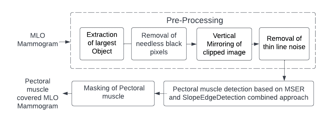
Breast Cancer is a fatal disease. Several people are losing their lives as a result of Breast Cancer. Mammography is the most often used Breast screening modality where we can see both mass and microcalcifications and both are the two major indicators of Breast Cancer. We can see Pectoral muscle also on MLO Mammograms. Digital Image Processing based computer aided diagnosis systems are being used widely to help the radiologist in detecting mass and microcalcifications in MLO Mammograms. However, because the intensity levels of the Pectoral muscle are similar to masses, in computer aided diagnosis system, Pectoral presence in the Mammogram has a detrimental effect on identifying mass. Therefore, in computer aided diagnosis system, Pectoral muscle masking substantially enhances lesion detection. This study suggests a novel ensemble computer aided diagnosis system strategy that combines the MSER based and SlopeEdgeDetection methods with extensive pre-processing to identify and cover Pectoral muscle from MLO Mammograms. The results demonstrate that the new procedure is straightforward and improves the precision of Pectoral region covering. Compared to the average accuracy of the state-of-the-art solutions which is 94%, the suggested technique achieves an accuracy of 99%. Performance analysis makes use of the Mini-MIAS database.
Article Details
References
https://my.clevelandclinic.org/health/diseases/2116 9-dense-Breast-tissue
Donoser, M. and Bischof, H. “Efficient Maximally Stable Extremal Region (MSER) Tracking”.
Woong Bae Yoon, Ji Eun Oh, Eun Young Chae, Hak Hee Kim, Soo Yeul Lee, Kwang Gi Kim, "Automatic Detection of Pectoral Muscle Region for Computer-Aided Diagnosis Using MIAS Mammograms", BioMed Research International, vol. 2016, Article ID 5967580, 6 pages, 2016. https://doi.org/10.1155/2016/5967580
P. S. Vikhe and V. R. Thool “Detection and Segmentation of Pectoral Muscle on MLO-View Mammogram Using Enhancement Filter”, (2017).
Samuel Rahimeto · Taye Girma Debelee · Dereje Yohannes · Friedhelm Schwenker "Automatic Pectoral muscle removal in mammograms", Springer 2019
"Geometry Based Pectoral Muscle Segmentation from MLO Mammogram Views", Saeid Asgari Taghanaki, IEEE Biomedical & Pharmacology Journal, September 2020.
Enas Mohammed Hussein Saeed, Hayder Adnan, "Pectoral Muscles Removal in Mammogram Image by Hybrid Bounding Box and Region Growing Algorithm ”, 2020.
G Vaira Suganthi, J Sutha, M Parvathy and C Durga Devi "Pectoral Muscle Segmentation in Mammograms", Biomed Pharmacol J 2020
"A Novel Approach to remove Pectoral muscle from Mediolateral Oblique Mammograms with Hybridization of MSERPectoral And HoughPectoral methods ", International Journal of Emerging Technologies and Innovative Research (www.jetir.org | UGC and issn Approved), ISSN:2349-5162, Vol.9,Issue 8,page no.ppf269-f276,August-2022,Available at: http://www.jetir.org/papers/JETIR2208526.pdf
https://web.ipac.caltech.edu/staff/fmasci/home/astro_refs/HoughTrans_lines_0 9.pdfctor-look- for-on-a-mammogram.html
"Robust Detection of Lines Using the Progressive Probabilistic Hough Transform", J. Matas, C. Galambos and J. Kittler
S. Suguna Mallika, D Rajya Lakshmi, “Mutation Testing and Its Analysis on Web Applications for Defect Prevention and Performance Improvement”, International Journal of e-Collaboration (IJeC) 17 (1), 71-88